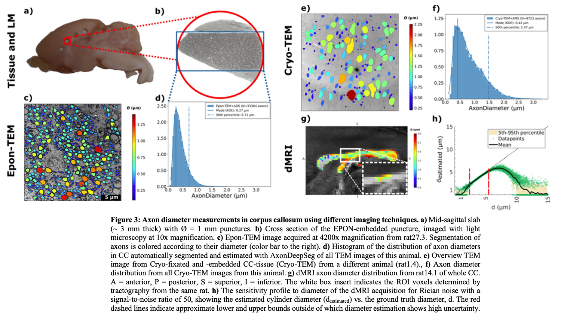
Abstract: "The structure-function relationship of myelinated nerve fibers (Waxman & Bennett, 1972) links axon diameter and myelin thickness, commonly expressed as the g-ratio, to conduction velocity via saltatory mechanisms. Here, we investigated this relationship in the transcallosal motor pathway of the rat brain by combining functional and structural metrics in the same animals. Transcallosal conduction times (TCTs) were measured using local field potentials (LFPs) evoked by optogenetic stimulation of excitatory neurons in the motor cortex. Conduction velocity estimates were obtained by combining TCTs with transcallosal tract lengths derived from diffusion MRI (dMRI)-based tractography. Fluorescent labeling of the viral optogenetic construct verified the tractography trajectories. In parallel, axon diameter and g-ratio were quantified using both dMRI and transmission electron microscopy (TEM). To assess dehydration-induced tissue shrinkage associated with conventional TEM preparation (“Epon-TEM”), we also performed cryo-fixation followed by TEM (“Cryo-TEM”) in a separate group. This revealed diameter-dependent axonal shrinkage, yielding a correction factor of 37%. Shrinkage correction improved agreement between dMRI and Epon-TEM estimates, although dMRI remained biased toward larger axons. When translated via the structure-function relationship, TCTs corresponded to smaller axons near the mode of the TEM diameter distribution, while dMRI-based diameters predicted TCTs that were too short compared with the recorded LFP latencies. Altogether, our findings show that structural and functional metrics differ in their sensitivity profiles. Accounting for such modality-dependent sensitivities facilitates the investigation of structure-function relationships, advancing our understanding of how microstructure supports neural communication."
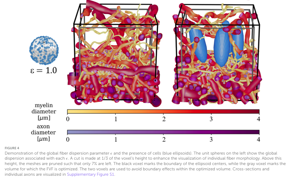
Abstract: "Brain white matter is a dynamic environment that continuously adapts and reorganizes in response to stimuli and pathological changes. Glial cells, especially, play a key role in tissue repair, inflammation modulation, and neural recovery. The movements of glial cells and changes in their concentrations can influence the surrounding axon morphology. We introduce the White Matter Generator (WMG) tool to enable the study of how axon morphology is influenced through such dynamical processes, and how this, in turn, influences the diffusion-weighted MRI signal. This is made possible by allowing interactive changes to the configuration of the phantom generation throughout the optimization process. The phantoms can consist of myelinated axons, unmyelinated axons, and cell clusters, separated by extra-cellular space. Due to morphological flexibility and computational advantages during the optimization, the tool uses ellipsoids as building blocks for all structures; chains of ellipsoids for axons, and individual ellipsoids for cell clusters. After optimization, the ellipsoid representation can be converted to a mesh representation which can be employed in Monte-Carlo diffusion simulations. This offers an effective method for evaluating tissue microstructure models for diffusion-weighted MRI in controlled bio-mimicking white matter environments. Hence, the WMG offers valuable insights into white matter's adaptive nature and implications for diffusion-weighted MRI microstructure models, and thereby holds the potential to advance clinical diagnosis, treatment, and rehabilitation strategies for various neurological disorders and injuries."
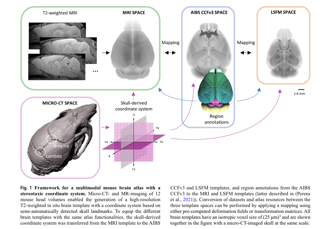
Abstract: "Magnetic resonance imaging (MRI) and light-sheet fluorescence microscopy (LSFM) are technologies that enable non-disruptive 3-dimensional imaging of whole mouse brains. A combination of complementary information from both modalities is desirable for studying neuroscience in general, disease progression and drug efficacy. Although both technologies rely on atlas mapping for quantitative analyses, the translation of LSFM recorded data to MRI templates has been complicated by the morphological changes inflicted by tissue clearing and the enormous size of the raw data sets. Consequently, there is an unmet need for tools that will facilitate fast and accurate translation of LSFM recorded brains to in vivo, non-distorted templates. In this study, we have developed a bidirectional multimodal atlas framework that includes brain templates based on both imaging modalities, region delineations from the Allen’s Common Coordinate Framework, and a skull-derived stereotaxic coordinate system. The framework also provides algorithms for bidirectional transformation of results obtained using either MR or LSFM (iDISCO cleared) mouse brain imaging while the coordinate system enables users to easily assign in vivo coordinates across the different brain templates."
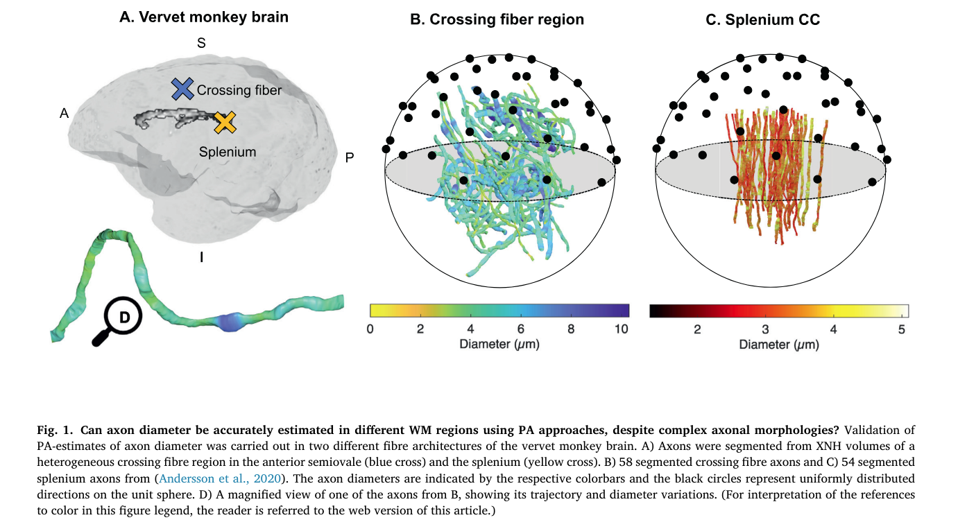
Abstract: "Noninvasive estimation of axon diameter with diffusion MRI holds the potential to investigate the dynamic properties of the brain network and pathology of neurodegenerative diseases. Recent studies use powder averaging to account for complex white matter architectures, but these have not been validated for real axonal geometries from regions that contain fibre crossings. Here, we present 120-304μm long segmented axons from X-ray nano-holotomography volumes of a splenium and crossing fibre region of a vervet monkey brain. We show that the axons in the complex crossing fibre region, which contains callosal, association, and corticospinal connections, exhibit a wider diameter distribution than those of the splenium region. To accurately estimate the axon diameter in these regions, therefore, sensitivity to a wide range of diameters is required. We demonstrate how the q-value, b-value, signal-to-noise ratio and the assumed intra-axonal parallel diffusivity influence the range of measurable diameters with powder average approaches. Furthermore, we show how Gaussian distributed noise results in a wider range of measurable diameter at high b-values than Rician distributed noise, even at high signal-to-noise ratios of 100. The number of gradient directions is also shown to impose a lower bound on measurable diameter. Our results indicate that axon diameter estimation can be performed with only few b-shells, and that additional shells do not improve the accuracy of the estimate. For strong gradients available on human Connectom and preclinical scanners, Monte Carlo simulations of diffusion confirm that powder averaging techniques succeed in providing accurate estimates of axon diameter across a range of diameters, sequence parameters and diffusion times, even in complex white matter architectures. At relatively low b-values, the diameter estimate becomes sensitive to axonal microdispersion and the intra-axonal parallel diffusivity shows time dependency at both in vivo and ex vivo intrinsic diffusivities."
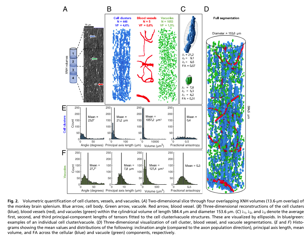
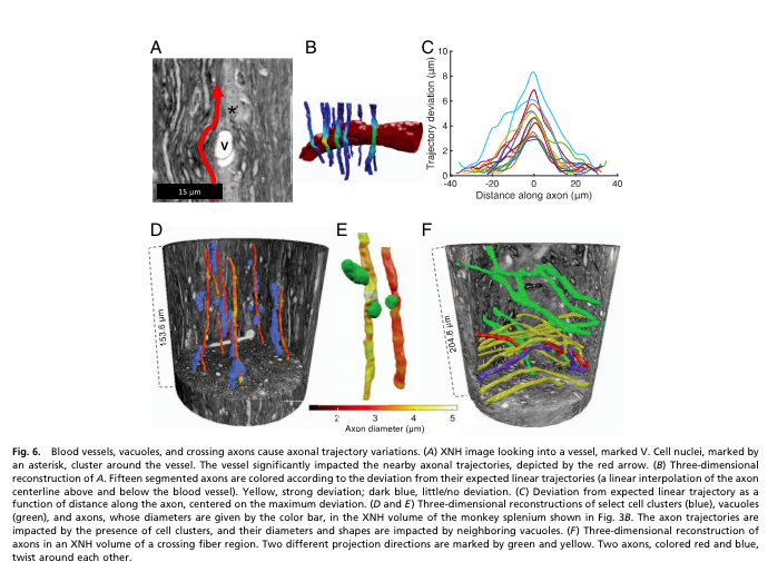
Abstract: "Axonal conduction velocity, which ensures efficient function of the brain network, is related to axon diameter. Noninvasive, in vivo axon diameter estimates can be made with diffusion magnetic resonance imaging, but the technique requires three-dimensional (3D) validation. Here, high-resolution, 3D synchrotron X-ray nanoholotomography images of white matter samples from the corpus callosum of a monkey brain reveal that blood vessels, cells, and vacuoles affect axonal diameter and trajectory. Within single axons, we find that the variation in diameter and conduction velocity correlates with the mean diameter, contesting the value of precise diameter determination in larger axons. These complex 3D axon morphologies drive previously reported 2D trends in axon diameter and g-ratio. Furthermore, we find that these morphologies bias the estimates of axon diameter with diffusion magnetic resonance imaging and, ultimately, impact the investigation and formulation of the axon structure-function relationship."
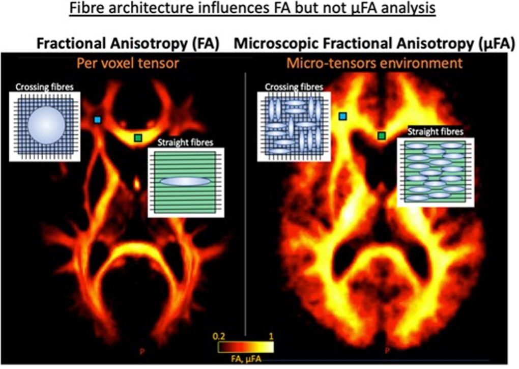

Abstract: "Multiple sclerosis leads to diffuse damage of the central nervous system, affecting also the normal-appearing white matter. Demyelination and axonal degeneration reduce regional fractional anisotropy in normal-appearing white matter, which can be routinely mapped with diffusion tensor imaging. However, the standard fractional anisotropy metric is also sensitive to physiological variations in orientation dispersion of white matter fibres. This complicates the detection of disease-related damage in large parts of cerebral white matter where microstructure physiologically displays a high degree of fibre dispersion. To resolve this ambiguity, we employed a novel tensor-valued encoding method for diffusion MRI, which yields a microscopic fractional anisotropy metric that is unaffected by regional variations in orientation dispersion. In 26 patients with relapsing-remitting multiple sclerosis, 14 patients with primary-progressive multiple sclerosis and 27 age-matched healthy controls, we compared standard fractional anisotropy mapping with the novel microscopic fractional anisotropy mapping method, focusing on normal-appearing white matter. Mean microscopic fractional anisotropy and standard fractional anisotropy of normal-appearing white matter were significantly reduced in both patient groups relative to healthy controls, but microscopic fractional anisotropy yielded a better reflection of disease-related white-matter alterations. The reduction in mean microscopic fractional anisotropy showed a significant positive linear relationship with physical disability, as reflected by the expanded disability status scale. Mean reduction of microscopic fractional anisotropy in normal-appearing white matter also scaled positively with individual cognitive dysfunction, as measured with the symbol digit modality test. Mean microscopic fractional anisotropy reduction in normal-appearing white matter also showed a positive relationship with total white-matter lesion load as well as lesion load in specific tract systems. None of these relationships between normal-appearing white-matter microstructure and clinical, cognitive or structural measures emerged when using mean fractional anisotropy. Together, the results provide converging evidence that microscopic fractional anisotropy mapping substantially advances the assessment of cerebral white matter in multiple sclerosis by disentangling microstructure damage from variations in physiological fibre orientation dispersion at the stage of data acquisition. Since tensor-valued encoding can be implemented in routine diffusion MRI, microscopic fractional anisotropy mapping bears considerable potential for the future assessment of disease progression in normal-appearing white matter in both relapsing-remitting and progressive forms of multiple sclerosis as well as other white-matter-related brain diseases."
We enable the estimation of the per-axon axial diffusivity from single encoding, strongly diffusion-weighted, pulsed gradient spin echo data. Additionally, we improve the estimation of the per-axon radial diffusivity compared to estimates based on spherical averaging. The use of strong diffusion weightings in magnetic resonance imaging (MRI) allows to approximate the signal in white matter as the sum of the contributions from only axons. At the same time, spherical averaging leads to a major simplification of the modeling by removing the need to explicitly account for the unknown distribution of axonal orientations. However, the spherically averaged signal acquired at strong diffusion weightings is not sensitive to the axial diffusivity, which cannot therefore be estimated although needed for modeling axons — especially in the context of multi-compartmental modeling. We introduce a new general method for the estimation of both the axial and radial axonal diffusivities at strong diffusion weightings based on kernel zonal modeling. The method could lead to estimates that are free from partial volume bias with gray matter or other isotropic compartments. The method is tested on publicly available data from the MGH Adult Diffusion Human Connectome project. We report reference values of axonal diffusivities based on 34 subjects, and derive estimates of axonal radii from only two shells. The estimation problem is also addressed from the angle of the required data preprocessing, the presence of biases related to modeling assumptions, current limitations, and future possibilities.
LinkIn magnetic resonance imaging, the application of a strong diffusion weighting suppresses the signal contribu tions from the less diffusion-restricted constituents of the brain’s white matter, thus enabling the estimation of the transverse relaxation time T2 that arises from the more diffusion-restricted constituents such as the axons. However, the presence of cell nuclei and vacuoles can confound the estimation of the axonal T2, as diffusion within those structures is also restricted, causing the corresponding signal to survive the strong diffusion weighting. We devise an estimator of the axonal T2 based on the directional spherical variance of the strongly diffusion-weighted signal. The spherical variance T2 estimates are insensitive to the presence of isotropic con tributions to the signal like those provided by cell nuclei and vacuoles. We show that with a strong diffusion weighting these estimates differ from those obtained using the directional spherical mean of the signal which contains both axonal and isotropically-restricted contributions. Our findings hint at the presence of an MRIvisible isotropically-restricted contribution to the signal in the white matter ex vivo fixed tissue (monkey) at 7T, and do not allow us to discard such a possibility also for in vivo human data collected with a clinical 3T system.
LinkDRCMR:
Nordre Hospitalsvej 13 2650 Hvidovre Captial Region of Denmark
DRCMR Main WebsiteContact: timd@drcmr.dk
DTU Compute:
Bygning 324 2800 Kongens Lyngby Captial Region of Denmark
DTU Compute Main WebsiteContact: tbdy@dtu.dk